
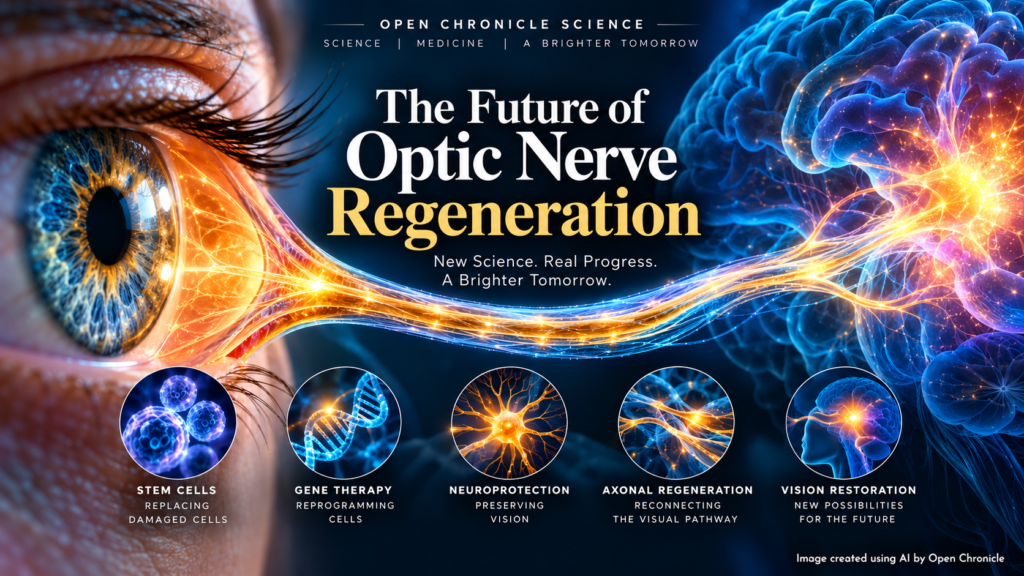
Gene therapy, epigenetic reprogramming, stem cells and neuronal regeneration are challenging one of neuroscience’s longest held assumptions: that damage to the optic nerve is permanent.
By Open Chronicle Science
For generations, damage to the optic nerve has carried a particularly difficult prognosis.
Once the retinal ganglion cells that connect the eye to the brain die, or their long nerve fibres are destroyed, medicine has had remarkably few ways of restoring what has been lost. Treatments for diseases such as glaucoma can reduce the risk of further damage. Doctors can lower intraocular pressure, control inflammation or address vascular and metabolic risk factors.
But rebuilding the connection between the eye and the brain has remained beyond the reach of medicine.
That assumption is now being challenged.
Across laboratories and early clinical trials, researchers are pursuing several radically different approaches to optic nerve repair. Some attempt to keep vulnerable neurons alive. Others try to rejuvenate damaged retinal cells. Still others seek to restart axonal growth or replace lost neurons entirely.
None has yet produced a clinically established method for rebuilding a severely damaged human optic nerve and restoring lost vision.
But something important has changed.
The question is increasingly moving from whether central nervous system neurons can ever regenerate to whether scientists can control that regeneration well enough to restore useful vision in humans.
The Optic Nerve Is Part of the Brain
Understanding the difficulty requires recognising what the optic nerve actually is.
It is not simply a cable connecting the eye to the brain.
The retina contains specialised neurons called retinal ganglion cells. These cells receive processed visual information from other retinal neurons and send it towards the brain through extremely long projections known as axons.
Approximately one million of these axons come together to form the human optic nerve.
They travel from the retina through the optic nerve, reach the optic chiasm and continue towards visual processing centres deeper inside the brain. From there, visual information ultimately reaches the visual cortex.
Damage can therefore occur at several levels.
A retinal ganglion cell may become dysfunctional but remain alive. Its axon may deteriorate. The cell may eventually die. Connections inside the brain may also be disrupted.
These stages are biologically very different.
Saving a stressed but living neuron is considerably easier than replacing a neuron that has disappeared.
That distinction lies at the centre of today’s research.
Three Different Battles
The emerging science of optic nerve restoration can broadly be divided into three objectives.
The first is neuroprotection: keeping retinal ganglion cells alive and functional for as long as possible.
The second is neuronal restoration: attempting to restore healthier function to cells that are damaged but still alive.
The third, and most ambitious, is true regeneration: replacing lost neurons or making surviving neurons grow new axons all the way back towards the appropriate regions of the brain.
Several experimental technologies are now attacking these problems simultaneously.
ER 100 and the Idea of Rejuvenating a Neuron
One of the most closely watched programmes in 2026 is ER 100, an experimental gene therapy being developed by Life Biosciences.
The therapy has entered a first in human Phase 1 clinical trial involving patients with open angle glaucoma and non arteritic anterior ischemic optic neuropathy, commonly known as NAION.
Its scientific concept is striking.
Rather than replacing retinal ganglion cells, ER 100 attempts to alter the biological state of cells that are still present.
The therapy uses an AAV2 viral vector to deliver three transcription factors known as OCT4, SOX2 and KLF4.
Together they are often referred to as OSK.
These factors are associated with cellular reprogramming, but the intention is not to convert retinal neurons into stem cells. Instead, researchers are investigating whether controlled partial epigenetic reprogramming can restore aspects of a younger cellular state without erasing the cell’s identity.
In simplified terms, the hypothesis is:
damaged or ageing retinal ganglion cell
↓
partial epigenetic reprogramming
↓
restoration of healthier gene expression
↓
improved neuronal function
↓
potential axonal repair or regeneration
Preclinical experiments have produced encouraging results in animal models, including work examining visual function and axonal architecture.
The crucial question, however, is whether any of this translates into humans.
October 2026 Could Provide an Important First Signal
That question is becoming particularly relevant now.
Life Biosciences announced on 1 October 2026 that it intends to present the first human data from the ongoing ER 100 Phase 1 trial at Eyecelerator during the American Academy of Ophthalmology meeting on 8 October.
The announcement should be interpreted cautiously.
First in human Phase 1 studies are primarily designed to investigate safety and tolerability. Early observations involving a small number of patients cannot establish that a treatment restores vision.
Nevertheless, the presentation represents an important milestone.
For the first time, researchers will begin to obtain direct evidence about what happens when this form of partial epigenetic reprogramming is attempted in the human eye.
If signs of biological or functional activity appear alongside acceptable safety, larger trials would still be required to determine whether the treatment provides meaningful clinical benefit.
If they do not, researchers will have learned something equally important about the limits of translating reprogramming biology from laboratory models into people.
CNTF: Keeping Neurons Alive
Another strategy is considerably less futuristic but potentially important.
Ciliary neurotrophic factor, or CNTF, is a protein capable of supporting neuronal survival.
An experimental implant known as NT 501 contains genetically engineered cells designed to continuously release CNTF inside the eye.
Instead of rebuilding an optic nerve, the objective is to protect retinal ganglion cells from degeneration and potentially improve the performance of neurons that remain viable.
Early clinical research in glaucoma has produced evidence of biological activity and supported further investigation in randomised studies.
The distinction is important.
A neuroprotective treatment could be extremely valuable even if it never regenerates a single lost axon.
Slowing or preventing retinal ganglion cell death could preserve useful vision for years or decades.
Could Metabolism Save the Optic Nerve?
Another increasingly important area concerns something much more fundamental: energy.
Retinal ganglion cells have demanding metabolic requirements. Their long axons require enormous amounts of energy to maintain electrical signalling and cellular transport.
Researchers increasingly suspect that metabolic dysfunction may contribute to the vulnerability of these neurons in diseases such as glaucoma.
That has led scientists to investigate nicotinamide, a form of vitamin B3 associated with cellular NAD metabolism.
Clinical studies have reported changes in retinal functional measurements following nicotinamide supplementation, while larger trials are investigating whether nicotinamide, sometimes combined with pyruvate, can produce meaningful benefits in glaucoma.
One major British study, NAMinG, recruited hundreds of participants to examine this possibility.
This does not mean that patients should begin taking very high doses of vitamin B3 themselves.
The quantities used in experimental research can be far higher than normal nutritional doses, and high dose nicotinamide can cause serious adverse effects, including liver toxicity.
The research therefore belongs in controlled clinical medicine, not self treatment.
Insulin as a Neuroprotective Treatment
Researchers are also investigating an unexpected candidate: insulin.
Insulin is best known for regulating blood glucose, but insulin signalling also influences neuronal survival and metabolism.
Clinical researchers are examining whether sterile insulin eye drops could have neuroprotective effects in glaucoma.
The TING2 study is designed to investigate this possibility, examining outcomes including retinal structure, visual fields, contrast sensitivity and visual acuity.
Again, the objective is primarily preservation and functional support rather than complete reconstruction of the optic nerve.
But preserving a vulnerable retinal ganglion cell today may prevent the need to replace it tomorrow.
Electrical Stimulation and the Brain’s Remaining Visual Networks
Not every experimental approach requires drugs or genes.
Researchers are also investigating whether electrical stimulation can improve the performance of damaged visual pathways.
One technique, repetitive transorbital alternating current stimulation, or rtACS, applies controlled electrical stimulation around the eyes.
The theory is that partially damaged visual systems may retain functioning neural networks that can be modulated or reorganised.
Clinical studies are investigating this approach in optic neuropathies and glaucoma.
Such stimulation would not resurrect dead neurons.
Instead, it raises another possibility: some visual impairment may involve neural networks that remain present but function inefficiently.
If so, part of future vision restoration could involve teaching surviving circuits to work better rather than physically rebuilding everything that has disappeared.
Stem Cells Change the Question Completely
Eventually, however, medicine encounters an unavoidable problem.
What happens when the retinal ganglion cell is already dead?
A dead neuron cannot be protected.
It cannot be metabolically strengthened.
And it cannot be rejuvenated.
It must somehow be replaced.
That is where stem cell research becomes particularly important.
Scientists can increasingly produce retinal neurons from pluripotent stem cells. The challenge is getting those new cells to behave like the neurons they are supposed to replace.
A transplanted retinal ganglion cell would need to survive inside the retina, integrate into the existing retinal circuitry and receive appropriate signals from neighbouring cells.
And that is only the beginning.
It would then have to grow an axon out of the retina and towards the brain.
Research at the Johns Hopkins Wilmer Eye Institute has recently addressed one of these obstacles by studying anatomical barriers that prevent transplanted retinal ganglion cells from integrating efficiently into retinal tissue.
Experimental work involving animal models, stem cells and donated human retinal tissue suggests that modifying these barriers can improve neuronal integration.
It is an important advance.
But successful integration into the retina still leaves an extraordinary journey ahead.
Restarting the Neuron’s Growth Programme
Another approach avoids replacing the neuron altogether.
What if scientists could convince an existing retinal ganglion cell to grow again?
During early development, neurons possess powerful growth programmes that allow their axons to travel enormous distances and locate appropriate targets.
Much of that regenerative capacity disappears as the nervous system matures.
Scientists are therefore searching for molecular switches capable of reactivating it.
One particularly interesting candidate is Nfe3.
Research supported by the US National Eye Institute has shown that increasing Nfe3 activity in experimental models can stimulate substantial axonal growth following optic nerve injury.
Conceptually, this offers an elegant solution.
Instead of building a replacement neuron, researchers would reactivate the regenerative machinery of the patient’s own surviving cell.
The neuron could then begin rebuilding its lost axon.
But growing an axon is only half the problem.
The Hardest Journey Is From the Eye to the Brain
Imagine that scientists successfully make thousands of retinal ganglion cells grow new axons.
Those axons cannot simply grow anywhere.
They must travel through the optic nerve.
They must reach the optic chiasm.
They must follow appropriate pathways.
They must reach the correct visual centres.
They must recognise the correct targets.
And ultimately they must form functional synapses capable of carrying meaningful visual information.
A connection that reaches the wrong region could be useless.
The challenge can therefore be represented as a biological sequence:
retinal ganglion cell
↓
axon regeneration
↓
optic nerve
↓
optic chiasm
↓
correct brain target
↓
synapse formation
↓
functional visual circuit
↓
useful vision
Every stage represents a major scientific problem.
This is why reports that scientists have “regrown the optic nerve” in an animal experiment need careful interpretation.
Growing axons beyond an injury is scientifically important.
It is not automatically equivalent to restoring normal sight.
Regeneration May Ultimately Require Combination Therapy
The most plausible future solution may therefore not be one miraculous treatment.
It may be several technologies used together.
A patient could theoretically receive neuroprotective treatment to prevent additional retinal ganglion cell death.
Epigenetic therapy might restore healthier function to damaged but surviving neurons.
Other molecular treatments could activate axonal growth.
Guidance molecules could help those axons navigate towards appropriate brain structures.
Electrical or other forms of neural stimulation might then encourage the reconstructed visual system to form and strengthen functional connections.
For advanced disease, stem cell derived retinal ganglion cells could eventually provide replacement neurons.
In other words, future optic nerve medicine may resemble reconstruction more than conventional drug treatment.
What Can Medicine Actually Do Today?
This is where expectations must remain realistic.
Modern ophthalmology can already do something enormously valuable: reduce the risk of additional optic nerve damage in many patients.
In glaucoma, lowering intraocular pressure remains fundamental.
Other optic neuropathies require treatment appropriate to their underlying cause.
What medicine cannot currently offer as routine clinical care is a treatment capable of reconstructing a severely destroyed human optic nerve and reliably restoring substantial vision that has been permanently lost.
There is no approved stem cell procedure that can rebuild the optic nerve.
There is no established gene therapy that makes a destroyed human optic nerve grow back to the brain.
There is no vitamin capable of regenerating millions of lost retinal ganglion cell axons.
Claims suggesting otherwise should therefore be treated with considerable caution.
But the Scientific Landscape Has Changed
That caution should not obscure the extraordinary change taking place.
A decade ago, optic nerve regeneration was overwhelmingly a problem of basic neuroscience.
Researchers were asking whether mature central nervous system neurons retained any meaningful capacity to regenerate.
Today the questions are becoming more precise.
Can the molecular brakes on axonal growth be removed?
Can an ageing neuron be epigenetically rejuvenated?
Can retinal ganglion cells derived from stem cells integrate into an adult human retina?
Can new axons be guided through the optic nerve?
Can regenerated axons identify their original targets?
Can the brain learn to interpret information arriving through a reconstructed pathway?
And, ultimately:
Can any of this restore useful sight to a human being?
For the first time, several pieces of that puzzle are being investigated simultaneously.
The Next Milestone
The immediate milestone will come on 8 October 2026, when the first human observations from the ER 100 programme are expected to be presented.
Those results should not be judged by whether researchers announce a dramatic restoration of sight.
At this stage, the more meaningful questions are simpler.
Was the therapy tolerated?
Were there serious adverse effects?
Did the treated retinal cells show measurable biological changes?
Were there objective changes in retinal or optic nerve function?
Did any visual measurements move in a direction that justifies a larger controlled trial?
If the answer to some of those questions is yes, it would still be only the beginning.
But it would represent another step across a boundary that neuroscience once regarded as extraordinarily difficult to cross.
The optic nerve remains one of the most challenging structures in the human nervous system to repair.
For the first time, however, scientists are attacking that challenge from almost every direction at once: metabolism, neuroprotection, gene therapy, epigenetics, electrical stimulation, axonal regeneration and stem cell biology.
Medicine cannot yet rebuild the connection between the eye and the brain.
The remarkable development of 2026 is that researchers are beginning to understand how it might one day be possible.
Sources and Further Reading
Clinical trials, research programmes and scientific institutions supporting this article.
ER 100 First in Human Phase 1 Study
Clinical trial registration covering experimental epigenetic gene therapy in open angle glaucoma and NAION.
ER 100 Optic Neuropathy Programme
Scientific and clinical information concerning the company’s partial epigenetic reprogramming programme.
First in Human ER 100 Data Presentation, October 2026
Announcement of the presentation of initial human data from the ongoing Phase 1 study at Eyecelerator 2026.
Seeing a Path to Nerve Regeneration
Research examining Nfe3 and the molecular mechanisms capable of stimulating retinal ganglion cell axonal regeneration.
Audacious Goals Initiative: Regenerating the Optic Nerve
Overview of neuronal survival, axonal regeneration, target selection, synapse formation and restoration of visual circuitry.
Translational Experiments Advance Efforts to Restore Vision With Transplanted Neurons
Research investigating how transplanted retinal neurons could overcome structural barriers to integration.
NAMinG Trial
Clinical investigation of nicotinamide in glaucoma and its potential role in retinal ganglion cell metabolism and neuroprotection.
This article describes experimental medical research and does not constitute medical advice.
Several therapies discussed remain investigational and are not approved treatments for optic nerve regeneration.
Patients with glaucoma, optic neuropathy or unexplained visual loss should discuss treatment decisions with an ophthalmologist or neuro ophthalmologist.