
OPEN CHRONICLE SCIENCE | NEUROSCIENCE · GENETICS · VISION RESEARCH
How Nobel Prize-winning optogenetics transformed a microscopic organism’s ability to sense light into a promising experimental treatment for vision loss.
By Open Chronicle Science Desk
October 2026 has brought two remarkable developments in neuroscience and vision research. On 5 October, the Nobel Prize in Physiology or Medicine was awarded to Karl Deisseroth, Peter Hegemann and Georg Nagel for their contributions to the development of optogenetics, a technology that allows scientists to control the activity of cells using light. Two days later, a clinical study published in the New England Journal of Medicine reported encouraging results from an experimental optogenetic treatment designed to restore aspects of visual function in people with advanced blindness.
The connection between these achievements reaches back more than two decades, to an organism that has no eyes, no brain and no nervous system.
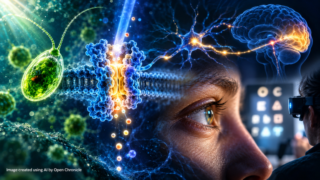
The single-celled green alga Chlamydomonas reinhardtii possesses a remarkable ability to detect light and adjust its movement accordingly. Scientists studying the molecular machinery behind this behaviour discovered proteins that could transform light into electrical signals.
Those proteins, known as channelrhodopsins, became the foundation of a scientific revolution.
By introducing genes encoding light-sensitive proteins into selected neurons, researchers developed a way to activate or inhibit neural circuits with extraordinary temporal precision. Optogenetics subsequently became one of the most influential experimental methods in modern neuroscience, helping scientists investigate how the brain processes information, generates behaviour and coordinates complex biological functions.
Today, the same underlying principle is being explored in ophthalmology.
For people whose retinal photoreceptors have been destroyed by degenerative disease, researchers are attempting to give surviving retinal cells a new ability to respond to light. Rather than replacing the original photoreceptors, these experimental therapies seek to create an alternative pathway through which visual information can reach the brain.
The results remain preliminary, and no optogenetic treatment can yet be described as a proven method for restoring normal human vision. Nevertheless, the research represents a striking example of how a discovery in fundamental biology can eventually inspire a medical intervention.
1. The Nobel That Illuminated Neuroscience
The 2026 Nobel Prize in Physiology or Medicine recognised a fundamental transformation in the way scientists investigate the nervous system.
For much of the twentieth century, neuroscientists relied on electrical stimulation, pharmacological interventions, recordings of neural activity and observations of patients with brain injuries to understand how neural circuits functioned.
These methods produced enormous advances, but they also had limitations.
Electrical stimulation could activate several types of neurons simultaneously. Drugs often affected broad populations of cells and operated over relatively long timescales. Recording neural activity revealed correlations between brain processes and behaviour, but establishing causal relationships remained difficult.
Optogenetics offered a different approach.
By genetically equipping particular cells with light-sensitive proteins, researchers could influence their electrical activity using carefully controlled illumination.
In suitable experimental preparations, this made it possible to activate selected neuronal populations within milliseconds, observe the resulting biological effects and investigate the contribution of specific circuits to behaviour.
The importance of the technology lies not simply in its ability to stimulate neurons, but in the combination of genetic targeting and precise optical control.
Scientists could begin asking questions that had previously been exceptionally difficult to investigate: Which neurons contribute to a particular memory? What happens when a specific neural pathway is activated? How do different circuits influence movement, fear, reward or perception?
The work recognised by the Nobel Prize brought together several scientific traditions, including microbial photobiology, molecular genetics, biophysics and systems neuroscience.
Its origins, however, were far removed from the human brain.
2. The Alga That Could Sense Light Without a Brain
Chlamydomonas reinhardtii is a microscopic green alga commonly found in freshwater environments.
Although it consists of a single cell, it possesses sophisticated mechanisms for responding to environmental conditions.
The organism contains chloroplasts that carry out photosynthesis and two flagella that enable it to swim through water.
It also has a specialized light-sensing apparatus commonly called an eyespot.
The eyespot is not an eye in the anatomical sense. It does not produce images, and the alga has no nervous system capable of interpreting visual scenes.
Instead, the eyespot and associated photoreceptor proteins help the cell detect light and regulate its swimming behaviour.
This capacity is known as phototaxis.
Depending on environmental conditions, the organism can move toward or away from light, allowing it to respond to conditions relevant to photosynthesis and survival.
For scientists, the intriguing question was how light detection could be translated into such a rapid behavioural response.
The answer involved proteins embedded in the cell membrane.
Research led by scientists including Peter Hegemann and Georg Nagel helped establish that certain algal photoreceptor proteins could function as light-gated ion channels.
These proteins were named channelrhodopsins.
Unlike many biological light-sensing systems that require several intermediate signalling steps, channelrhodopsins can directly couple light absorption to the opening of an ion channel.
This property would prove extraordinarily valuable.
A protein evolved to help a microscopic organism navigate its environment could potentially be used to control electrical activity in completely different types of cells.
3. From Channelrhodopsins to the Control of Neurons
The key molecular breakthrough emerged in the early 2000s.
In 2002 and 2003, researchers published influential studies identifying and characterising channelrhodopsins from Chlamydomonas.
One particularly important protein was Channelrhodopsin-2, or ChR2.
ChR2 contains a light-sensitive retinal chromophore, a molecule derived from vitamin A. When the protein absorbs light of an appropriate wavelength, it undergoes a structural change that opens an ion-conducting pathway across the cell membrane.
Positively charged ions can then flow through the channel, changing the electrical potential of the membrane.
In neurons, this change can trigger an action potential — the electrical impulse through which nerve cells communicate.
The implications were immediate.
If the gene encoding ChR2 could be introduced into a neuron and the protein expressed in its membrane, light could become a means of controlling neuronal firing.
In 2005, Edward Boyden, Feng Zhang, Ernst Bamberg, Georg Nagel and Karl Deisseroth published a landmark study in Nature Neuroscience demonstrating precisely this possibility in mammalian neurons.
The researchers showed that light could drive neural activity with millisecond-scale precision.
This was a decisive step in the development of modern optogenetics.
Subsequent work expanded the range of available proteins, enabling scientists not only to activate neurons but also to inhibit or otherwise modulate their activity.
Different microbial opsins offered distinct properties, including sensitivity to different wavelengths of light, varied response speeds and different effects on membrane electrical activity.
The result was an increasingly versatile molecular toolkit.
4. How Optogenetics Works
Optogenetics combines two central technologies: genetic modification and optical stimulation.
The genetic component determines which cells become sensitive to light.
The optical component determines when and where those cells are stimulated.
In a typical research application, scientists introduce genetic instructions for a light-sensitive protein into selected cells, often using a modified viral vector.
The cells then manufacture the protein and incorporate it into their membranes.
When researchers illuminate the cells with the appropriate wavelength of light, the protein changes its activity.
Depending on the type of opsin used, the resulting ion movement can increase or decrease the probability that a neuron will generate electrical impulses.
This process allows researchers to manipulate neural circuits in a highly controlled manner.
Importantly, the technology does not mean that scientists can read thoughts or directly reconstruct complex mental experiences.
Neural circuits contain many interacting cell types, and behaviour emerges from coordinated activity across distributed networks.
Optogenetics provides a powerful experimental tool for investigating these networks, but understanding the complete relationship between neural activity and conscious experience remains a major scientific challenge.
Nevertheless, the technology has transformed experimental neuroscience.
Researchers have used it to investigate neural pathways associated with movement, learning, memory, sensory processing, sleep and emotional behaviour.
It has also contributed to research into neurological and psychiatric conditions, although translating experimental findings into clinical treatments is considerably more difficult than demonstrating effects in laboratory models.
5. A Revolution in Brain Research
One of optogenetics’ greatest scientific contributions has been its ability to help researchers distinguish correlation from causation.
Before the technology became widely available, scientists could identify neurons that became active during particular behaviours, but determining whether those neurons were necessary or sufficient for the behaviour was often difficult.
Optogenetics made it possible to manipulate defined neural populations and observe the consequences.
In animal studies, researchers have investigated how particular circuits contribute to fear responses, reward-related behaviour, locomotion and memory.
Other experiments have examined neural pathways involved in sleep regulation and the processing of sensory information.
The technology has also been combined with advanced imaging techniques, electrophysiological recordings and computational analysis.
Together, these methods have helped researchers build increasingly detailed descriptions of how neuronal networks operate.
However, findings from experimental animals do not automatically translate into treatments for humans.
Human brains contain highly complex networks, and therapeutic applications require careful evaluation of safety, delivery methods, long-term effects and ethical implications.
The most immediate medical applications of optogenetics may therefore emerge in tissues where light can be delivered relatively directly.
The retina is one such tissue.
6. The Human Retina: Where Light Becomes Information
Human vision begins when light enters the eye and reaches the retina, a thin layer of neural tissue lining the back of the eyeball.
The retina is not merely a passive light detector.
It is a sophisticated biological processing system that transforms incoming light into electrical signals and begins analysing visual information before it reaches the brain.
Two major types of photoreceptors perform the initial light-detection process.
Rods are highly sensitive to low levels of illumination and play an essential role in night vision.
Cones support colour vision and high-resolution visual perception, particularly under brighter lighting conditions.
When light reaches these cells, specialised photopigments initiate biochemical reactions that change their electrical activity.
The resulting signals are processed through networks of bipolar cells, horizontal cells, amacrine cells and retinal ganglion cells.
The axons of retinal ganglion cells form the optic nerve, which carries visual information toward the brain.
Visual perception depends on the coordinated operation of this entire system.
It also depends on the brain’s ability to interpret patterns of neural activity as shapes, movement, colour, contrast and spatial relationships.
When retinal photoreceptors are destroyed, the first stage of this processing chain is severely disrupted.
Yet some downstream retinal neurons may remain alive.
This observation is central to the development of optogenetic therapies for blindness.
7. When Photoreceptors Die: Understanding Retinitis Pigmentosa
Retinitis pigmentosa refers to a diverse group of inherited retinal disorders that cause progressive degeneration of photoreceptor cells.
The condition can arise from mutations in numerous genes involved in retinal structure and function.
Symptoms often begin with difficulties seeing in low light.
As the disease progresses, affected individuals may experience narrowing of peripheral vision, reduced visual acuity and, in advanced cases, profound visual impairment or blindness.
The precise course varies considerably between patients and genetic forms of the disease.
In many cases, rods are affected before cones, although the underlying mechanisms differ between disorders.
For people with advanced retinal degeneration, the loss of photoreceptors creates a major therapeutic challenge.
Conventional approaches that depend on functioning photoreceptors may no longer be effective.
Gene replacement therapies can be valuable for certain inherited retinal diseases, but they are not universally applicable.
Some treatments require viable photoreceptors and target specific genetic mutations.
Optogenetics proposes a different strategy.
Instead of correcting the original genetic defect or regenerating the missing photoreceptors, researchers attempt to make surviving retinal neurons directly sensitive to light.
The objective is to bypass damaged parts of the visual pathway.
This approach could potentially be relevant to patients with different genetic causes of retinal degeneration, provided that enough downstream retinal circuitry remains functional.
It is a compelling idea, but it also involves substantial biological compromises.
Retinal ganglion cells were not designed to function as conventional photoreceptors.
They normally receive signals that have already undergone extensive processing.
Making them directly light-sensitive may allow some visual information to reach the brain, but it cannot automatically reproduce the sophisticated encoding performed by a healthy retina.
8. Teaching the Retina to Respond to Light Again
One of the best-known clinical approaches to optogenetic vision restoration involves the protein ChrimsonR.
ChrimsonR is a light-sensitive microbial opsin that responds to wavelengths extending into the orange-red region of the visible spectrum.
Researchers have investigated whether the protein can be expressed in surviving retinal ganglion cells in people with advanced photoreceptor degeneration.
The treatment uses an engineered adeno-associated viral vector, or AAV, to deliver genetic instructions for the protein into the eye.
Following administration, the targeted cells may begin producing the light-sensitive protein.
However, ordinary environmental light may not provide the intensity or characteristics needed to stimulate the modified cells effectively.
For this reason, one experimental system combines gene delivery with specialised electronic goggles.
A camera captures information from the surrounding environment, while an electronic processor converts the images into patterns of light suitable for activating the introduced opsin.
The goggles therefore perform part of the function that would ordinarily be carried out by the eye’s natural optical and retinal systems.
The modified retinal cells respond to these optical signals, potentially transmitting information through the optic nerve to the brain.
This creates an artificial visual pathway.
It is not equivalent to replacing the retina or restoring its original structure.
Instead, the system attempts to provide a new form of sensory input that the brain may learn to interpret.
9. The First Major Clinical Milestone: 2021
In May 2021, a research team led by José-Alain Sahel and collaborators published an important study in Nature Medicine.
The report described a patient with advanced retinitis pigmentosa who received an optogenetic treatment combining an AAV vector encoding ChrimsonR with specialised light-stimulating goggles.
The patient had experienced severe visual impairment for many years.
Following treatment and training with the goggles, he demonstrated an ability to perceive, locate and interact with certain objects under controlled experimental conditions.
The researchers also recorded brain activity associated with visually guided tasks.
The results provided evidence that optogenetic stimulation could produce functionally useful visual signals in a human participant with advanced retinal degeneration.
This was a landmark proof of concept.
For the first time, an approach developed from microbial light-sensitive proteins had demonstrated the potential to restore limited aspects of visual function in a person with severe blindness.
The achievement was scientifically significant, but its limitations were equally important.
The study described one participant, and the observed visual abilities were restricted.
The treatment did not restore normal sight, reading vision or conventional colour perception.
Performance depended on the experimental system, including the specialised goggles and the participant’s adaptation to the new visual signals.
Nevertheless, the results justified continued investigation.
10. October 2026: A New Chapter in Clinical Research
The publication of new clinical findings in October 2026 represents another important stage in the development of optogenetic vision restoration.
The study, Optogenetic Therapy for Restoring Aspects of Visual Function, published in the New England Journal of Medicine, reported results from ten participants with advanced retinitis pigmentosa who received an experimental treatment involving ChrimsonR.
The research was conducted as a phase I clinical trial, with safety as its principal objective.
The investigators also assessed changes in visual function.
Seven participants demonstrated increased light sensitivity, while six showed improvements that met the study’s criteria for clinically meaningful visual change.
Among participants who experienced improvements in light sensitivity, the reported increases ranged from approximately twofold to more than sixtyfold.
These findings suggest that the treatment may produce measurable functional changes in some individuals with severe retinal degeneration.
However, the results must be interpreted cautiously.
The study included a small number of participants and did not have a randomised control group.
Visual improvement was not uniform, and increased sensitivity to light is not equivalent to restoration of normal vision.
The trial also recorded ocular adverse events, including a serious episode of transient central retinal artery occlusion.
Although that event resolved rapidly, it illustrates why safety assessment remains essential when evaluating experimental ocular gene therapies.
The study therefore represents encouraging early evidence, not definitive proof of clinical effectiveness.
Larger controlled trials and longer follow-up will be necessary to determine which patients may benefit, how durable the effects are and whether the potential advantages outweigh the risks.
11. Other Experimental Approaches to Vision Restoration
The ChrimsonR strategy is not the only form of optogenetic therapy under investigation.
Researchers are exploring different microbial opsins, retinal cell targets and methods of delivering visual stimulation.
One approach involves multi-characteristic opsins designed to respond to a broader range of light conditions.
The experimental therapy MCO-010, developed by Nanoscope Therapeutics, uses an optogenetic protein intended to confer light sensitivity on surviving retinal neurons.
Unlike systems that rely on specialised electronic goggles, this approach seeks to produce useful responses under ambient lighting conditions.
Clinical investigations, including the RESTORE and REMAIN programmes, have reported findings concerning visual acuity, functional vision and the persistence of treatment effects.
However, differences in trial design, patient populations, measurement methods and therapeutic mechanisms make direct comparisons between experimental treatments difficult.
The field also faces a broader question: which retinal cells should be targeted?
Ganglion cells offer a relatively direct route to the optic nerve, but activating them bypasses much of the retina’s normal processing machinery.
Targeting bipolar cells could potentially preserve more of the retina’s internal circuitry, although gene delivery and reliable expression present additional challenges.
Researchers are also investigating ways to improve light sensitivity, reduce the intensity of illumination required and develop proteins with more favourable biological properties.
The ultimate objective is not simply to produce a measurable response to light.
It is to restore visual information that is sufficiently detailed, stable and useful to improve everyday activities.
12. The Scientific Challenges That Remain
Despite the progress achieved, several obstacles stand between experimental optogenetic therapies and widespread clinical application.
Reproducing the complexity of natural vision
Healthy human vision depends on millions of photoreceptors and complex retinal circuits.
These circuits extract information about contrast, movement, colour and spatial patterns before transmitting signals to the brain.
Optogenetic stimulation of a smaller population of surviving cells cannot automatically reproduce this processing.
Delivering genes safely and effectively
Viral vectors must deliver genetic material to the intended cells without causing unacceptable inflammation or other complications.
The distribution of gene expression may vary between patients.
Researchers must also establish whether the introduced proteins remain functional over long periods.
Achieving sufficient light sensitivity
Some microbial opsins require stronger illumination than natural photoreceptors.
Electronic devices may help overcome this limitation, but they add complexity and can affect the practicality of treatment.
Understanding brain adaptation
People who have experienced severe blindness for many years may need time and training to interpret unfamiliar visual signals.
The brain’s capacity to adapt is important, but it varies between individuals and is not unlimited.
Measuring meaningful improvements
A laboratory test showing increased sensitivity to light does not necessarily demonstrate that a patient can navigate independently, recognise faces or read.
Future trials must therefore assess functional outcomes that matter in everyday life.
Establishing long-term safety
Gene-based interventions may produce effects lasting for years.
This potential durability is attractive, but it also means that unexpected long-term effects must be carefully investigated.
These challenges explain why promising early clinical results should not be confused with an established cure.
13. From Fundamental Biology to Translational Medicine
The history of optogenetics illustrates one of the most important principles of scientific progress: discoveries made without an immediate medical objective can eventually transform clinical research.
Scientists studying a microscopic alga were initially attempting to understand how a single cell detects and responds to light.
They were not developing a treatment for blindness.
The identification of channelrhodopsins created a new possibility in molecular biology.
Neuroscientists recognised that these proteins could be introduced into neurons and used to control electrical activity.
Further advances in genetic engineering, optical instrumentation and experimental neuroscience transformed the idea into a widely used research technology.
Ophthalmologists and vision scientists subsequently explored whether the same molecular principles could help patients whose retinal photoreceptors had been lost.
The resulting clinical programmes brought together specialists in molecular genetics, neuroscience, retinal biology, ophthalmology, medical devices and rehabilitation.
This progression was neither immediate nor inevitable.
It required decades of research, repeated experimental validation and contributions from many scientific disciplines.
The Nobel Prize recognises a foundational part of that journey.
The clinical studies demonstrate how the technology is now being tested in an entirely different context.
14. The Future: Can Optogenetics Restore Useful Vision?
The next stage of optogenetic vision research will depend on larger clinical trials, improvements in molecular engineering and a clearer understanding of which patients are most likely to benefit.
Scientists are working to develop proteins that respond more efficiently to light, targeting methods that improve cellular specificity and devices capable of delivering more useful visual information.
Advances in image processing may also contribute.
For treatments involving external cameras and optical stimulation, computational systems could potentially emphasise relevant features of the environment, such as object boundaries, movement or contrast.
However, these improvements would need to be evaluated experimentally rather than assumed to produce better vision.
Another major objective is to determine how well patients can learn to interpret the artificial visual signals generated by optogenetic systems.
Rehabilitation and training may prove essential components of treatment.
Future clinical research must also establish whether the benefits persist, whether repeated intervention is necessary and how the therapies compare with other approaches to retinal degeneration.
These include gene-specific treatments, retinal prostheses, stem-cell-based strategies and other forms of regenerative medicine.
Each approach addresses a different aspect of the biological problem.
It is therefore unlikely that a single technology will be appropriate for every cause or stage of blindness.
Optogenetics may ultimately become one component of a broader therapeutic landscape.
Conclusion: The Long Journey from an Alga to Human Vision
The story of optogenetics begins with a deceptively simple biological question.
How does a microscopic organism without a brain know where the light is?
The search for an answer revealed proteins capable of converting light into changes in electrical activity.
Those proteins allowed scientists to develop a new way of studying the nervous system, transforming experimental neuroscience and opening opportunities to investigate the biological foundations of behaviour.
The same molecular machinery is now being explored as a possible treatment for some forms of blindness.
The clinical evidence remains preliminary. Patients receiving experimental optogenetic therapies have not regained normal vision, and important questions concerning safety, effectiveness and long-term outcomes remain unresolved.
Yet the scientific achievement is already substantial.
Researchers have demonstrated that surviving retinal neurons can be made responsive to light and that, under certain conditions, this can provide limited but measurable visual information to people with advanced retinal degeneration.
The significance of the work extends beyond ophthalmology.
It illustrates how basic biological research can generate tools whose eventual applications are impossible to predict at the moment of discovery.
A protein that evolved to help an alga navigate toward light became a method for investigating the brain.
That method is now helping scientists explore whether light can once again carry useful information to a visual system damaged by disease.
The journey from a single-celled organism to experimental human therapy is far from complete.
But it has already changed the way scientists understand the relationship between light, neurons and perception.
And it offers a compelling reminder that some of medicine’s most promising possibilities begin with curiosity about the natural world.
Scientific Sources and Further Reading
Primary research, peer-reviewed publications and scientific institutions documenting the development of optogenetics and its emerging applications in vision restoration.
The Nobel Prize in Physiology or Medicine 2026
Official Nobel Prize materials concerning the development of optogenetics and the contributions of Karl Deisseroth, Peter Hegemann and Georg Nagel.
Channelrhodopsin-2, a Directly Light-Gated Cation-Selective Membrane Channel
Nagel, G., et al. (2003). Proceedings of the National Academy of Sciences. A foundational study identifying the light-gated ion channel properties of Channelrhodopsin-2, which later became central to the development of optogenetics.
Millisecond-Timescale, Genetically Targeted Optical Control of Neural Activity
Boyden, E. S., Zhang, F., Bamberg, E., Nagel, G., and Deisseroth, K. (2005). Nature Neuroscience, 8, 1263–1268. Landmark experimental work demonstrating that genetically introduced light-sensitive proteins could control neuronal activity with millisecond precision.
Optogenetics
Deisseroth, K. (2011). Nature Methods, 8, 26–29. A scientific review examining the principles, development and applications of optogenetic technology in experimental neuroscience.
Partial Recovery of Visual Function in a Blind Patient After Optogenetic Therapy
Sahel, J.-A., et al. (2021). Nature Medicine, 27, 1223–1229. A pioneering clinical report describing partial recovery of visual function in a patient with advanced retinitis pigmentosa following experimental optogenetic treatment combined with specialised light-stimulating goggles.
Optogenetic Therapy for Restoring Aspects of Visual Function
Sahel, J.-A., et al. (2026). New England Journal of Medicine, 395, 1399–1408. Clinical research investigating the safety and preliminary functional outcomes of an experimental optogenetic treatment for individuals with advanced retinal degeneration.
European Society of Cataract and Refractive Surgeons
Clinical developments involving MCO-010 optogenetic therapy for vision restoration. Coverage of emerging treatment strategies, clinical findings and potential applications in patients with advanced retinal degeneration.
Editorial Note
This article is based on peer-reviewed scientific publications, historical research and clinical findings available as of 18 October 2026. Optogenetic therapies for vision restoration remain investigational. Reported improvements in light sensitivity or selected visual tasks should not be interpreted as restoration of normal sight or evidence of an established cure for blindness. The Open Chronicle Science Desk distinguishes established molecular mechanisms from preliminary clinical outcomes and will update this article as further evidence becomes available.