
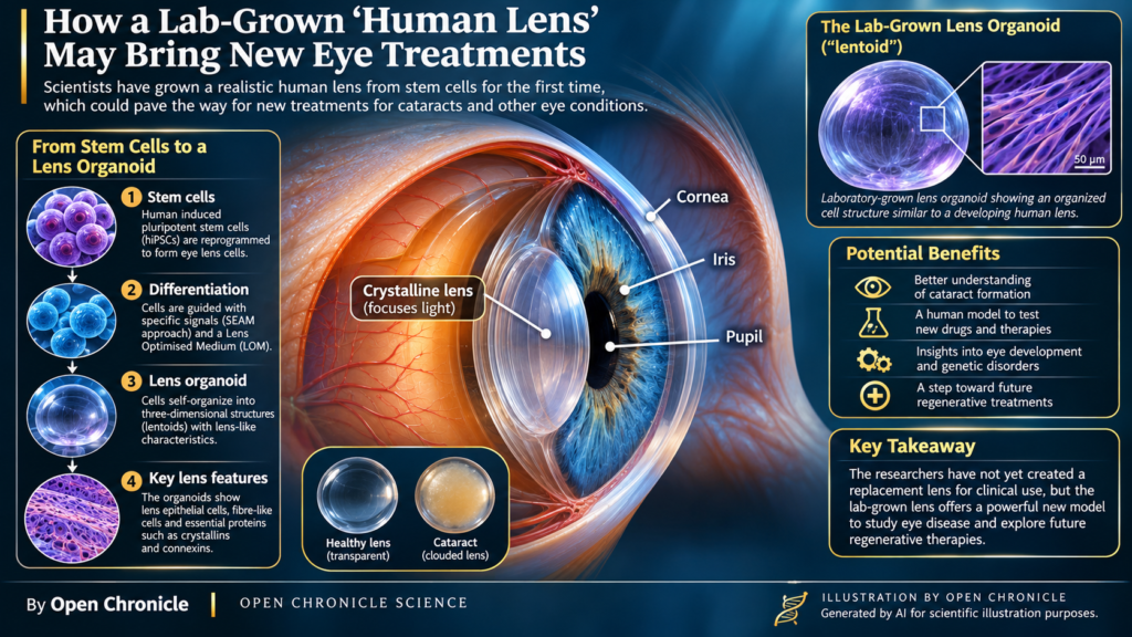
An international research team has recreated key features of the developing human eye lens in the laboratory, offering a sophisticated new model for investigating cataracts, cellular differentiation and the possibilities of regenerative ophthalmology.
By Open Chronicle
The human eye contains one of the body’s most remarkable biological structures: a transparent, flexible lens capable of focusing light with extraordinary precision. Throughout much of our lives, it quietly adjusts its optical properties, allowing us to shift our gaze between distant landscapes and the smallest details of the world around us.
Yet this sophisticated structure is also vulnerable. As the lens ages, its proteins can lose the molecular organization that keeps it transparent. Light begins to scatter rather than pass cleanly through, vision becomes increasingly clouded, and cataracts develop.
For millions of people, the solution is surgery, removing the clouded natural lens and replacing it with an artificial intraocular lens.
But what if scientists could investigate the development, deterioration and potential regeneration of the human lens without relying exclusively on donated tissue or animal models?
A new study published in npj Regenerative Medicine brings researchers closer to answering that question.
An international team from Anglia Ruskin University, Cardiff University and the University of Osaka has developed three dimensional human lens organoids from induced pluripotent stem cells. These laboratory grown structures reproduce several important features of the developing human crystalline lens, including specialized cell populations, structural proteins and elements of its intricate tissue architecture.
The achievement does not mean that scientists have created a fully functional replacement lens ready for transplantation. Instead, it provides something potentially just as valuable for the immediate future: a biologically sophisticated experimental system in which researchers can observe how human lens tissue forms, organizes itself and responds to its environment.
And that could transform how scientists investigate one of the world’s leading causes of visual impairment.
The extraordinary biology of a transparent lens
To understand the importance of this discovery, it is necessary to appreciate just how unusual the human crystalline lens really is.
Located behind the iris and pupil, the lens works alongside the cornea to focus incoming light onto the retina, where photoreceptor cells convert photons into electrical signals that the brain interprets as vision.
Unlike most tissues, however, the mature lens must remain almost perfectly transparent while maintaining a highly ordered internal structure.
Its transparency depends on several biological adaptations.
The lens contains densely packed, elongated fibre cells filled with specialized proteins known as crystallins. These proteins are arranged in ways that minimize fluctuations in refractive index and reduce the scattering of visible light.
During maturation, lens fibre cells undergo a remarkable transformation. They progressively eliminate their nuclei and other intracellular organelles, reducing structures that would otherwise interfere with the passage of light.
The resulting tissue combines optical transparency with mechanical flexibility.
THE HUMAN CRYSTALLINE LENS
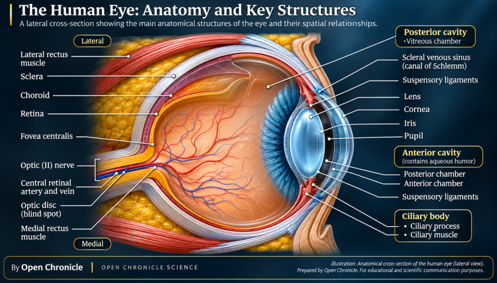
Another essential component is the lens capsule, a specialized basement membrane composed of extracellular matrix proteins. This capsule surrounds the lens, helps maintain its shape and provides mechanical support during accommodation, the process through which the eye changes focus.
The lens also has no direct blood supply. Instead, it relies on a carefully regulated internal circulation system involving water, ions and metabolites.
Gap junction proteins, particularly connexins, allow neighbouring cells to exchange small molecules and maintain the physiological conditions required for transparency.
Any significant disruption to these systems can compromise optical performance.
This is why reproducing the lens in a laboratory is considerably more complicated than simply growing a cluster of human cells.
Scientists must recreate aspects of the developmental programme that instructs cells where to position themselves, which proteins to express and how to establish the architecture of a functioning optical tissue.
Reprogramming human cells to recreate eye development
The researchers began with human induced pluripotent stem cells, commonly abbreviated as hiPSCs.
These cells are produced by reprogramming differentiated adult cells into a pluripotent state, restoring their capacity to generate many different specialized cell types.
The development of induced pluripotency fundamentally changed regenerative biology because it allowed scientists to obtain versatile human cell populations without relying exclusively on embryonic stem cells.
However, pluripotency alone is not sufficient to produce an organized tissue.
Cells must receive a carefully coordinated sequence of biochemical and environmental signals that resembles the developmental instructions operating inside an embryo.
In the case of the eye, this process is particularly complex.
During embryogenesis, the crystalline lens originates from the surface ectoderm, a layer of embryonic tissue that interacts with the developing optic vesicle.
These interactions induce the formation of the lens placode, a thickened region of cells that subsequently invaginates to form the lens vesicle.
As development progresses, cells within the vesicle acquire different identities. Anterior cells contribute to the lens epithelium, while posterior cells elongate and differentiate into primary lens fibres.
Further growth depends on the continued proliferation and differentiation of epithelial cells near the lens equator.
The new research attempts to reproduce important elements of this developmental sequence under controlled laboratory conditions.
The SEAM approach: recreating developmental organization
A central component of the experimental strategy was a differentiation system known as SEAM, short for self formed ectodermal autonomous multi zone.
This approach encourages pluripotent stem cells to organize into distinct regions containing cell populations associated with different ectodermal developmental pathways.
Rather than forcing every cell into an identical state, the method exploits the capacity of developing cells to respond collectively to molecular signals and spatial relationships.
That distinction matters.
Embryonic development is not simply a process in which individual cells acquire specialized identities. It is also a coordinated process of morphogenesis, in which cells change shape, migrate, interact and assemble into organized anatomical structures.
By using a system that reproduces aspects of this spatial organization, the researchers sought to establish conditions favourable to lens differentiation.
They also developed and used a specialized culture formulation, referred to as Lens Optimised Medium, or LOM, to support the development and maturation of lens like structures.
The resulting organoids exhibited characteristics associated with different stages of lens formation.
This provided the research team with an opportunity to investigate not only which cells were present, but also how those cells organized themselves into increasingly complex tissue.
Inside the laboratory grown lens
One of the most significant findings was the emergence of structures containing distinct cellular features associated with the natural lens.
The organoids displayed epithelial regions alongside elongated cells resembling differentiating lens fibres.
This is an important distinction because a biological lens is not merely a transparent sphere. It contains specialized cellular compartments with different functions.
The anterior lens epithelium maintains cellular activity and contributes to growth and tissue homeostasis.
Lens fibre cells, by contrast, undergo extensive elongation and differentiation, accumulating proteins required for optical function.
The investigators examined the laboratory grown structures using molecular characterization and advanced microscopy.
Their observations included the expression of lens associated crystallins, connexins and proteins involved in cellular adhesion.
These molecular markers are relevant because they provide evidence that the cells were following developmental programmes associated with lens identity rather than forming an unrelated tissue aggregate.
Crystallins: the proteins that make transparency possible
Among the most important molecules identified were crystallins.
These proteins constitute a major proportion of the soluble protein content of the mature lens.
The α-crystallins, which belong to the small heat shock protein family, contribute to protein stability and help protect other lens proteins from aggregation.
β-crystallins and γ-crystallins provide structural and optical properties that are essential to the highly concentrated protein environment of lens fibre cells.
Their arrangement influences the refractive index of the tissue and its ability to transmit light without excessive scattering.
The expression of crystallins in laboratory grown lens organoids therefore provides an important indicator of lens specific differentiation.
Nevertheless, the presence of crystallins does not automatically establish that an organoid possesses the full optical performance of a natural human lens.
Optical transparency, refractive power, elasticity and long term protein stability are separate properties that require direct functional evaluation.
That distinction will be crucial as the technology develops.
Connexins and the communication network inside the lens
The researchers also examined proteins associated with intercellular communication.
Connexins form channels that connect adjacent cells through gap junctions.
In the lens, these channels are essential because the tissue lacks conventional blood vessels and must distribute ions and metabolites through an internal network of cellular communication.
Connexin 46 and connexin 50, encoded by GJA3 and GJA8 respectively, are particularly important in lens physiology.
Mutations affecting these proteins have been associated with inherited cataracts and other abnormalities of lens development.
The identification of connexin related features in lens organoids is therefore relevant not only to developmental biology but also to the investigation of genetic eye disorders.
A sufficiently mature organoid model could eventually allow researchers to examine how specific mutations alter cellular communication, tissue organization and optical transparency.
Seeing cellular architecture at microscopic resolution
The study went beyond identifying proteins.
Using ultrastructural analysis, the researchers examined the physical organization of cells within the developing organoids.
Electron microscopy revealed elongated cellular structures and specialized patterns of contact between neighbouring cells.
Among the features described were interdigitations resembling ball and socket junctions.
These structures are associated with the complex interfaces between natural lens fibre cells.
In a mature lens, fibre cells are arranged in tightly packed layers, and their membranes contain specialized structures that help maintain mechanical cohesion and tissue organization.
The appearance of comparable features in laboratory grown tissue suggests that the cells were reproducing aspects of lens morphogenesis at a structural level.
This is an important achievement because many organoid systems reproduce molecular markers of a tissue without fully recreating its anatomical complexity.
The ability to generate recognizable tissue architecture brings the model closer to the biological environment researchers wish to investigate.
However, resemblance at the microscopic level should not be confused with complete physiological equivalence.
A laboratory organoid may reproduce selected developmental features while still lacking the precise geometry, mechanical properties or long term homeostatic mechanisms of an adult human lens.
The molecular instructions behind lens formation
The development of the crystalline lens is regulated by an intricate network of transcription factors and signalling pathways.
Several of these regulatory systems are particularly relevant to understanding the new research.
PAX6 is widely recognized as a central regulator of eye development. It participates in the establishment of ocular identity and influences multiple stages of lens formation.
SOX2 and SIX3 also contribute to early developmental processes associated with ocular and lens specification.
As differentiation progresses, additional regulators become important.
PROX1 plays a role in lens fibre cell differentiation, while SOX1 is associated with aspects of lens development and cellular maturation.
These transcription factors do not operate independently.
They participate in regulatory networks influenced by signalling molecules, including fibroblast growth factors, bone morphogenetic proteins and components of the Wnt signalling system.
The concentration, timing and spatial distribution of these signals help determine whether cells remain proliferative, adopt an epithelial identity or differentiate into elongated lens fibres.
Understanding these interactions is essential for regenerative medicine.
If scientists can determine which molecular signals govern each developmental transition, they may eventually gain greater control over the production of specific lens cell populations.
That knowledge could improve experimental models, support drug discovery and inform future strategies for tissue repair.
A surprising discovery: lens cells capable of generating new organoids
Perhaps one of the most intriguing aspects of the research concerns the developmental potential retained by certain epithelial like cells.
The team identified populations expressing PAX6, SIX3 and SOX2, molecular markers associated with early ocular and lens development.
These cells exhibited self renewing characteristics and were capable of forming secondary lentoids under appropriate experimental conditions.
The subsequent activation of transcription factors including PROX1 and SOX1 was associated with further lens differentiation.
This observation suggests that some cells within the laboratory system retained a degree of developmental plasticity.
In biological terms, plasticity describes the capacity of cells to alter their differentiation state or developmental behaviour in response to specific signals.
For regenerative medicine, this is particularly interesting.
One of the major obstacles to repairing complex tissues is identifying cell populations capable of generating the required specialized structures while preserving appropriate organization and function.
The formation of secondary lentoids suggests that the experimental system could help researchers investigate how lens progenitor populations are maintained, activated and directed toward differentiation.
It does not demonstrate that the adult human lens can regenerate spontaneously after injury.
Nevertheless, it offers a controlled environment in which the molecular mechanisms underlying regenerative potential can be examined.
Cataracts: a global challenge that extends beyond ageing
The potential importance of this research becomes clearer when considered alongside the burden of cataract disease.
Cataracts develop when the normally transparent crystalline lens becomes progressively opaque.
Although ageing is the most common contributing factor, cataracts can also result from genetic mutations, metabolic disorders, ocular trauma, inflammation and prolonged exposure to certain medications.
At the molecular level, cataract formation frequently involves changes in protein stability and organization.
Crystallins are unusually long lived proteins. In the central regions of the adult lens, many remain present for decades because mature fibre cells have lost the machinery required for conventional protein renewal.
Over time, these proteins can undergo oxidation, deamidation, truncation and other post translational modifications.
Such changes may alter their solubility, promote aggregation and increase the scattering of light.
The lens also depends on antioxidant defence systems, including glutathione metabolism, to protect its proteins from oxidative damage.
When these protective mechanisms become insufficient, the accumulation of damaged proteins can contribute to progressive opacification.
Why a laboratory model could change cataract research
Traditional cataract research has relied on combinations of animal experiments, isolated human lens tissue, cell cultures and biochemical studies.
Each approach has limitations.
Animal lenses differ from human lenses in aspects of development, ageing and physiology. Human tissue is difficult to obtain at precisely defined developmental stages. Conventional cell cultures often lack the three dimensional organization of intact tissue.
Human lens organoids could help address some of these limitations.
By generating lens associated structures from pluripotent stem cells, researchers may be able to examine disease mechanisms within a more developmentally relevant human cellular environment.
For example, future experiments could investigate how inherited mutations affect crystallin expression, how oxidative stress changes cellular organization or how candidate compounds influence protein aggregation.
Patient derived hiPSCs could also make it possible to investigate particular genetic forms of cataract disease.
Such models would not automatically reproduce the decades long ageing processes that contribute to common age related cataracts. Developing experimental systems that adequately simulate those processes remains an important challenge.
Nevertheless, the new platform offers an additional way to study the relationship between molecular dysfunction and lens pathology.
Could scientists eventually replace cataract surgery with regeneration?
For now, cataract surgery remains the established and highly effective treatment for clinically significant lens opacity.
During the procedure, the clouded natural lens material is removed, usually using phacoemulsification, and an artificial intraocular lens is implanted.
Modern surgical techniques have transformed the treatment of cataracts, allowing many patients to recover useful vision through a relatively short procedure.
However, artificial lenses do not reproduce every physiological property of the natural crystalline lens.
In particular, restoring the full dynamic accommodation of a young human eye remains a challenge.
A natural lens changes its shape in response to forces transmitted through the zonular fibres and ciliary muscle.
Most conventional intraocular lenses provide a relatively fixed optical configuration, although multifocal, extended depth of focus and accommodating designs seek to address different aspects of postoperative visual performance.
A genuinely regenerated biological lens would therefore represent a different therapeutic approach.
Instead of replacing the natural lens with an artificial optical implant, regenerative strategies would aim to restore or reconstruct living tissue with appropriate transparency, geometry and mechanical function.
Yet that objective remains scientifically demanding.
A successful biological replacement would need to maintain long term transparency, achieve appropriate refractive properties, integrate mechanically with surrounding ocular structures and remain stable throughout the patient’s lifetime.
The 2026 organoid study does not establish that these requirements have been achieved.
Its immediate contribution is to improve the experimental understanding of lens development, not to provide a clinically validated alternative to cataract surgery.
The earlier experiments that made this possible
The concept of lens regeneration did not begin with the latest study.
In 2016, researchers reported an experimental surgical approach designed to preserve endogenous lens epithelial stem and progenitor cells during treatment of congenital cataracts in young children.
The study provided evidence that retained cells could contribute to the formation of new lens tissue under specific clinical circumstances.
Although promising, the findings did not establish a broadly applicable treatment for adult cataracts.
The biological environment of an infant lens differs considerably from that of an ageing adult lens, particularly in terms of cellular activity, tissue growth and regenerative potential.
Other investigations subsequently explored the generation of lens epithelial cells and microlenses from human pluripotent stem cells.
These studies helped demonstrate that human cells could be directed toward lens associated developmental programmes outside the body.
The latest research builds upon this scientific foundation by emphasizing spatial organization, tissue morphology, molecular differentiation and epithelial plasticity.
Its significance lies not in the simple existence of laboratory grown lens like structures, which had been demonstrated previously, but in the combination of developmental characteristics reproduced within the new experimental platform.
Beyond cataracts: a new tool for developmental biology
Although cataract research represents an obvious potential application, the broader scientific value of the organoids may extend well beyond a single disease.
The eye is a powerful model for studying embryonic development because its formation depends on precisely coordinated interactions between different tissues.
Lens induction, in particular, requires communication between the developing optic structures and surrounding ectoderm.
These interactions involve transcriptional regulation, growth factor signalling, changes in cell adhesion and extensive morphological reorganization.
Laboratory systems capable of reproducing selected stages of this process may help scientists investigate fundamental questions about human organogenesis.
How do initially similar cells acquire distinct developmental identities?
How do local signalling gradients influence tissue organization?
What determines whether an epithelial cell remains proliferative or begins terminal differentiation?
And how do developing tissues coordinate molecular identity with physical architecture?
These questions are relevant not only to ophthalmology but also to the wider fields of developmental biology and tissue engineering.
The same general principles of cellular differentiation, extracellular matrix organization and morphogenesis operate across numerous organs.
Understanding them in a relatively accessible experimental model could therefore contribute to broader regenerative research.
The obstacles between laboratory discovery and clinical treatment
Despite its promise, the new platform faces several scientific and technological challenges before any therapeutic application could be considered.
One major question concerns reproducibility.
Organoids frequently exhibit biological variability, with differences in size, cellular composition, maturation and structural organization between individual cultures.
For a model to become widely useful in pharmaceutical development or disease research, scientists must establish reliable production methods and standardized criteria for evaluating tissue quality.
Another challenge is functional maturation.
Expressing the molecular markers of a developing lens is not equivalent to reproducing the optical and mechanical performance of a mature crystalline lens.
Future research will need to examine properties such as transparency, refractive behaviour, elasticity and long term cellular stability.
Protein homeostasis represents another major obstacle.
The natural lens maintains an exceptionally concentrated protein environment over many decades.
Reproducing this degree of stability in engineered tissue may require a much deeper understanding of crystallin regulation, proteostasis and cellular stress responses.
There are also safety considerations associated with pluripotent stem cell technologies.
Any future transplantation strategy would require rigorous evaluation of residual undifferentiated cells, abnormal proliferation, genetic stability, immune compatibility and integration with surrounding tissues.
These are not minor technical details. They are fundamental requirements for translating laboratory discoveries into safe clinical interventions.
A new perspective on the future of vision
The most important consequence of the research may ultimately be a change in how scientists investigate the human lens.
For generations, cataracts have largely been approached as a problem of removing and replacing damaged optical tissue.
Regenerative biology introduces a complementary question: can researchers understand the developmental instructions that create the lens well enough to reproduce, preserve or potentially restore its biological properties?
The answer remains incomplete.
But the ability to generate human lens organoids with recognizable epithelial populations, specialized structural proteins and features of developing fibre cell architecture represents meaningful progress.
It gives scientists a new experimental system in which to examine the processes that make the lens transparent, the molecular disruptions that cause disease and the developmental mechanisms that may one day inform regenerative strategies.
The human crystalline lens is a small structure, but its biology is extraordinarily complex.
By recreating selected elements of that complexity in the laboratory, researchers have opened a new window into the science of vision.
And while a laboratory grown replacement lens remains a distant possibility rather than an established treatment, the discoveries made along the way could reshape how scientists understand, investigate and eventually treat diseases of the human eye.
Research Publication
Journal
npj Regenerative Medicine
Article
Human iPSC-derived lens organoids recapitulate lens development and morphogenesis in vitro
Authors
Jodie Harrington, Robert D. Young, Xiaoqin Li,
Ryuhei Hayashi, Justyn W. Regini,
Andrew J. Quantock, Barbara Pierscionek
and Kohji Nishida.
Published
15 October 2026
Digital Object Identifier (DOI)
10.1038/s41536-026-00514-6
Sources and Further Reading
npj Regenerative Medicine | Original Research Article
The primary peer reviewed investigation describing the
production, cellular differentiation, molecular
characterization and structural organization of human
lens organoids.
Cardiff University | Institutional Research Repository
The university’s academic record of the investigation,
including access to the published research.
American Academy of Ophthalmology | Lens Regeneration Research
Background on earlier stem cell research and experimental
approaches to lens regeneration.
PubMed | Human Lens Cell and Microlens Research
Earlier research investigating the production of
lens related cells and microlenses from human
pluripotent stem cells.
Editorial Note
This article is an independent scientific feature prepared
by Open Chronicle using published research and established
ophthalmological knowledge. The experimental results concern
laboratory grown lens organoids and do not demonstrate a
clinically approved regenerative treatment for cataracts.
Therapeutic applications discussed in this article remain
prospective and require further investigation.